An Introduction To Double Outlet Right Ventricle (DORV)
Congenital heart defects (CHDs) are a class of structural flaws that impair the heart’s early development during pregnancy. An uncommon disorder that affects the regular flow of blood through the heart is the Double Outlet Right Ventricle (DORV), which is one of these complicated CHDs. We shall examine the complexities of DORV in this article, including its etiology, symptoms, diagnosis, and available treatments.
Recognising DORV
The aorta and the pulmonary artery, the two major blood arteries that deliver oxygen-rich and oxygen-poor blood, respectively. They start from the right ventricle instead of their customary locations in people with DORV, a congenital heart abnormality. The aorta and pulmonary artery emerge from the left and right ventricles, respectively, of a healthy heart. As a result, the aorta and pulmonary artery both exit the right ventricle in DORV, which can result in a number of problems.

Reasons for DORV
Although the precise origin of DORV is yet unknown, genetic and environmental factors are thought to have a role. The development of this illness may also be influenced by genetic abnormalities, maternal health during pregnancy, and exposure to certain chemicals. DORV might furthermore happen as part of more complex heart abnormalities in some cases.
Symptoms
The symptoms of DORV can vary depending on the specific anatomy of the heart and the degree of blood mixing between the two circulatory systems. Common symptoms may include:
- Cyanosis: Babies with DORV often have bluish skin and lips due to inadequate oxygen supply to the body.
- Rapid Breathing and Fatigue: Infants may exhibit rapid breathing and tire easily during feeding or physical activity.
- Poor Growth: Some infants with DORV may experience slow growth and weight gain due to the increased effort required to pump blood.
- Heart Murmur: A heart murmur, an abnormal sound that a turbulent blood flow produces, is often present and undergoes detection during a physical examination.
Diagnosis
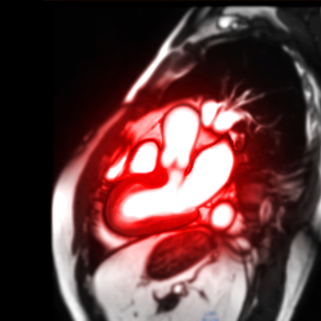
Prenatal screening, physical examinations, and imaging studies are commonly used to diagnose DORV. Regular ultrasound exams throughout pregnancy can occasionally detect the problem. During a physical examination after delivery, a paediatrician could find heart murmurs or other problematic indications. Confirmatory exams including echocardiography, cardiac catheterization, and MRI aid in supplying a more thorough understanding of the anatomy and operation of the heart.
Classification of DORV
Depending on the precise configuration of the great arteries and the existence of other cardiac abnormalities, DORV can manifest in a variety of ways. The following are the two main categories:
- Subaortic Ventricular Septal Defect (VSD): This kind of DORV has a VSD. It is a hole between the two lower chambers of the heart, that rests beneath the aorta. This can result in blood that is both oxygen-rich and oxygen-poor, which can worsen the symptoms.
- Subpulmonary VSD: In this kind of VSD, the VSD is situated below the pulmonary artery. Even though it is still complicated, this type of DORV may cause fewer severe symptoms. This is because of a more even blood flow.
Therapy Alternatives
The determination of the method of treating DORV is totally upon the patient’s general health, related problems, and particular anatomical characteristics. The objectives of treatment are to increase oxygenation, lessen symptoms, and avoid consequences. Possible course of action includes:
- Surgery: To correct DORV, surgery is frequently the only option. Depending on the precise types of cardiac abnormalities present, several types of surgery take place. The aorta may need redirection, VSDs may need sealing, and normal blood flow patterns may need restoration.
- Palliative treatments: Palliative treatments are very useful in some situations. Particularly, when surgery is not immediately feasible, to increase oxygenation and reduce symptoms. These operations could involve expanding heart holes already present or creating a shunt to channel blood.
- Medication: Medication may be a recommendation to treat symptoms and improve cardiac performance. Inotropes, diuretics, and antiarrhythmic medications are a common prescription .
- Ongoing Monitoring: To track the child’s development, modify medicines, and determine whether additional therapies are necessary, regular follow-up meetings with a paediatric cardiologist are crucial.
Prognosis
Thanks to developments in medical and surgical procedures, the prognosis for people with DORV has greatly improved recently. For a positive outcome, early diagnosis and adequate treatment are essential. Many children with DORV can live healthy lives and develop normally with the right treatment. However, periodic procedures and lifetime cardiac monitoring is extremely essential.
Thus,
There is an impact on the normal flow of blood through the heart by the complicated congenital heart. This is an abnormality mostly doctors term as Double Outlet Right Ventricle. Early identification and improvements in medical and surgical therapies significantly improve prognosis for each and every dorv patient. This is despite the fact that it poses unique obstacles. Many children with DORV can live happy lives and reach their full potential with continuing care and supervision. To guarantee the greatest results for persons living with this illness, parents and other carers must collaborate closely with medical professionals.
For more information or surgery related queries, get in touch with us at Travocure.


Hey, cool post You can check if there’s a problem with your website with Internet Explorer. Because of this issue, many readers will overlook your excellent writing because IE is still the most popular browser.
1